posterior part of Norma basalis
Introduction :
Norma basalis is the basal view of the skull. considering it as one of the most difficult parts of studying the skull, the following short notes will help you revise and get through it with ease.
Anatomical divisions:
For the sake of understanding, Norma basalis is divided into three parts
1.Anterior part of Norma basalis
2. Middle part of Norma basalis
3. posterior part of Norma basalis
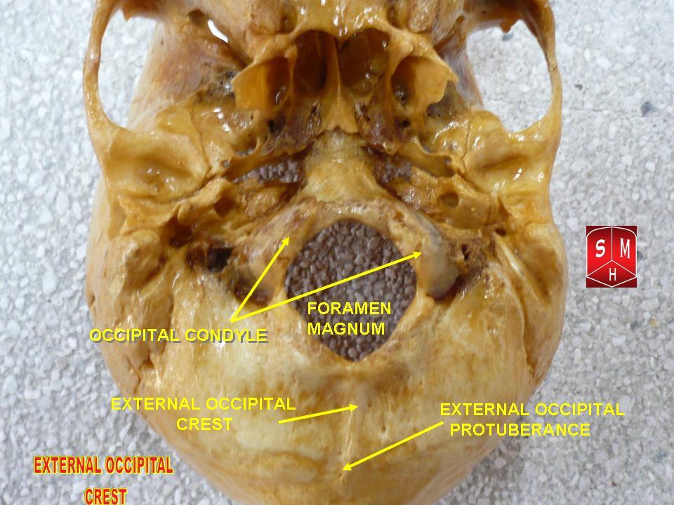
Posterior part of norma basalis
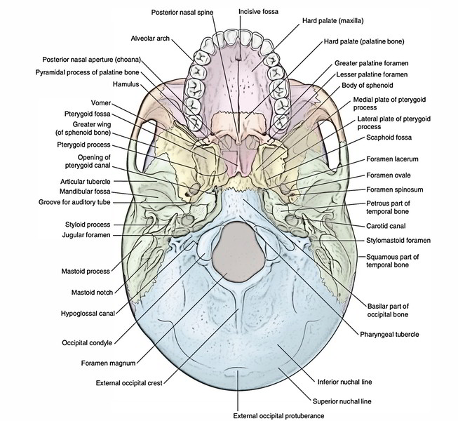
contents of posterior part:
Foramen:
- foramen magnum
- hypoglossal canal
- posterior condylar canal
- stylomastoid foramen
- jugular foramen
Processes :
- External occipital crest
- condylar part of occipital bone
- styloid process
- occipital condyle
- pharyngeal tubercle
- highest nuchal line
- superior nuchal line
- inferior nuchal line
Foramen magnum
– divided into small anterior and large posterior compartments using alar ligaments of axis vertebra:

Anterior compartment:
- Apical ligament of dens.
- Upper longitudinal band of cruciform ligament of the atlas.
- Membrana tectoria: a continuation of posterior longitudinal ligament of vertebral bodies.
Posterior compartment:
- Medulla oblongata and its meninges.
- Two posterior spinal a.s (right and left).
- Anterior spinal a.
- Communicating veins between internal vertebral venous plexus and basilar venous plexus.
- Two vertebral a.s (right and left).
- Sym. plexus around vertebral a.s.
- Spinal roots of two accessory nerves (right and left).
Hypoglossal canal
- The twelfth cranial (hypoglossal) nerve.
- Meningeal branch of the ascending pharyngeal artery.
Jugular foramen
- inferior petrosal sinus
- sigmoid sinus
- the glossopharyngeal nerve ( 9th cranial nerve)
- Tenth cranial (vagus) nerve.
- eleventh cranial nerve(accessory nerve)
11th and 10th cranial nerve are surrounded by cranial sheet whereas the 9th cranial nerve has an independent sheet of dura matter
Posterior condylar canal
- Transmits emissary vein
- connects sigmoid sinus with suboccipital venous plexus
Stylomastoid foramen
- facial nerve
- stylomastoid branch of posterior auricular nerve
Processes:
external occipital crest :
Condylar part of occipital bone
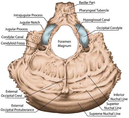
The condylar parts are located lateral to the foramen magnum. They comprise two kidney-shaped prominences (occipital condyles) that articulate with the first cervical vertebra (atlanto-occipital joint).
the hypoglossal nerve pierces the condylar process after coming out of the hypoglossal canal
STYLOID PROCESS
present in front of the mastoid process
Foramen:
stylomastoid foramen: transmits stylomastoid artery and facial nerve
Muscular attachments:
- stylomandibular ligament
- styloglossus muscle
- stylophyrangeus muscle
- stylohyoid muscle
- stylohyoid ligament

Phyrangeal tubercle:
muscular attachment of pharyngeal tubercle :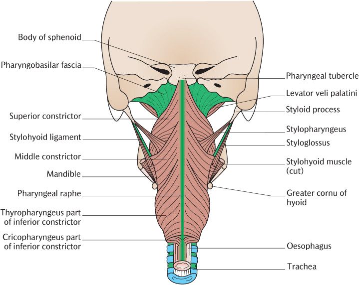
- superior constrictor
- rectus capitus anterior
- longus capitus
- supports pharyngeal tonsil
nuchal lines:

Highest nuchal line:
attached to the epicranial aponeurosis
Superior nuchal line:
- trapezius
- sternocleidomastoid
- splenius capitus
- occipital belly of occipitofrontalis
Inferior nuchal line:
above superior nuchal line:
- semispinalis capitis
below inferior nuchal line:
- rectus capitus posterior major
- rectus capitus posterior minor
- obliques superior


Comments
Post a Comment