Anatomy of middle ear
INTRODUCTION
The ear is an organ of hearing. It is also concerned with maintaining the equilibrium of the body One hears with the ears. The centre for hearing is in the temporal lobe of the brain above the ear
It consists of three parts:
- External ear
- Middle ear
- Internal ear
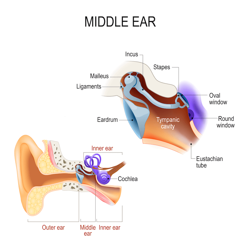
MIDDLE EAR
it is also called the tympanic cavity or tympanum
the middle ear is a narrow air-filled cavity situated in the petrous part of the temporal bone, between the external and internal ear
shape and size:
the middle ear is shaped like a cube with a middle biconcave cavity
the distance between lateral wall are
- 6 mm nea the roof
- 2mm in the center
- 4 mm in the floor

communications:
- anteriorly with the nasopharynx through the auditory tube
- posteriorly with the mastoid antrum and the mastoid air cells
BOUNDARIES
Tegmental wall or roof :
- the middle ear is separated from the middle cranial fossa by a thin plate of bone called tegmen tympani
- in young children, the roof has a gap between the unossified petrous temporal
jugular wall or floor :
- it is made of a thin plate of the temporal bone.
- tympanic canaliculus transmits the tympanic branch of glossopharyngeal nerve


carotid wall or anterior wall:
- bears opening for tensor tympani
- opening of auditory tube
- it separates the middle ear from the internal carotid canal
- processus cochleariformis
- the boney septum between tensor tympani and auditory tubule
Mastoid wall or posterior wall:
- aditus
- opening through which the epitympanic recess communicates with the mastoid antrum
- fossa includes
- depression which lodges the incus
- pyramid
- conical projection that has an opening for the tendon of stapedius muscle
- posterior cannaliculus for chorda tympani
- chorda tympani enters the middle ear.
Membranous wall or latereal wall:
- separates middle ear from the external acoustic meatus
- it is formed by two components
- tympanic membrane
- squamous temporal bone
- it contains two apertures
- petrotympanic fissure
- lodges anterior process of malleus
- transmits tympanic branch of maxillary artery
- anterior cannaliculus
- transmits chorda tympani
- it leaves middle ear through this to emerge at the base of the skull
Labyrinth or medial wall :
- promontory
- rounded bulging produced by first turn of cochlea
- fenestra cochlea
- oval opening posterosuperior to the promontory
- fenestra cochlea
- opening to the posteroinferior of the promontory
- the prominence of facial canal
- runs backwards to reach the margin of audits
- prominance of lateral semilunalr canal
- sinus tympani
- depression behind promontory
CONTENTS
the middle ear contains the following
- bones
- malleus
- incus
- stapes
- joint between ossicles
- incudomalleolar joint
- incudostapedial joint
- muscles
- tensor tympani
- stapedius
- vessels
- nerves
- chorda tympani
- tympanic plexus
- Air
Malleus:
the word malleus means hammer in Latin.
it is the largest and most laterally placed ossicle

It contains 5 parts
- head
- lies in the epitympanic recess
- provides attachment to superior and lateral ligaments
- neck
- lies against the pars flaccida
- related medially to chorda tympani
- Anterior process
- connected to the petrotympanic fissure
- lateral process
- provides attachment to the superior and lateral ligaments
- handle
- it is attached to the upper half of tympanic membrane
Incus :
it is so-called because it resembles an anvil
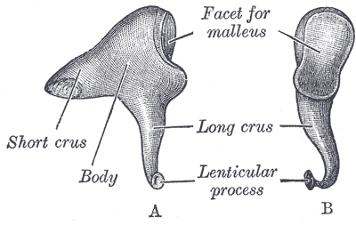
it has 2 parts :
- body
- bears articulating surface that articulates with the head of malleus
- long process
- projects downwards
- bears lentiform nodule which articulates with head of the stapes
Stapes :
the smallest and most medially placed bone in the ear

it has the following parts :
- head
- has concave surface that articulates with the lentiform nodule
- neck
- provides posterior insertion to the stapedius muscle
- two limbs
- short and less curved anteriorly
- the posterior part is long and attached to the footplate
- footplate
- oval shape
- fits in fenester vestibuli
Muscles of the middle ear :
middle ear contains two muscle tensor tympani and the stapedius
both muscles work together to reduce the intensity of sound
- Tensor tympani

- arises and lies in the boney canal
- it ens in a tendon which passes laterally to insert into the handle of the malleus
- it is supplied by mandibular nerve
- Stapedius

- les in the boney canal related to the posterior wall
- the muscle arises in the boney canal and gets inserted into the neck of the stapes
arterial supply :
1. anterior tympanic branch
2. posterior tympanic branch of stylomastoid
3. posterior and superior tympanic branches of middle meningeal artery
4. branches of ascending pharyngeal artery
5. tympanic branch of internal carotid artery
venous drainage: superior petrosal sinus and pterygoid plexus
lymphatic drainage: preauricular and retropharyngeal lymph nodes
nerve supply:
1. tympanic branch of glossophayrengeal nerve
- supplies the mucous membrane of middle ear, auditory tube, mastoid antrum and air cells
2. superior and inferior carotiotympanic nerves
- vasomotor to the mucous membrane
FUNCTIONS OF MIDDLE EAR
middle ear transmits sound waves from the external ear to the internal ear.the intensity of sound is increased 10 times by ossicles

Comments
Post a Comment