Anatomy of larynx
INTRODUCTION
the larynx is the organ for the production of voice. it is also an air passage and acts as a sphincter for the inlet of the respiratory passage.
POSITION
larynx lies in the anterior midline of the neck extending from the root of the tongue
SIZE
length - 44mm in males and 36 mm in females
at puberty male larynx grows larger, resulting in a louder voice and low pitched
the growth of the female larynx is negligible therefore the voice is high pitched
FEATURES
the larynx is made up of a framework of cartilage
they are connected by joints, ligaments and membrane
Their movements are controlled by several muscles
CARTILAGES OF LARYNX
the larynx contains 9 unique cartilage with 3 paired and 3 unpaired cartilage
Unpaired cartilage :
2. cricoid cartilage
3. epiglottis

Paired cartilage :
1. Arytenoid cartilage

2. corniculate cartilage 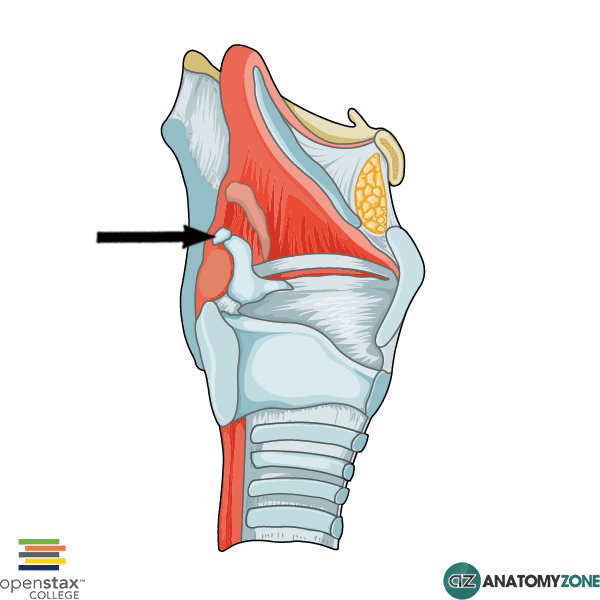
3. cuniform cartilage

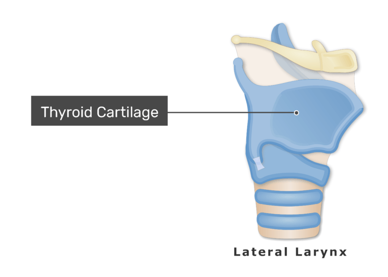
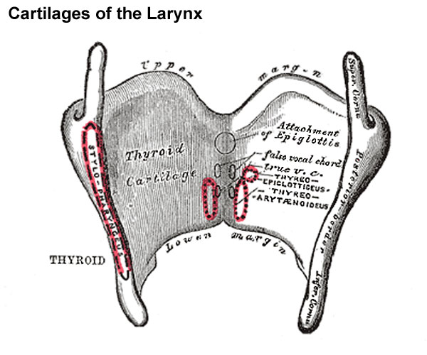
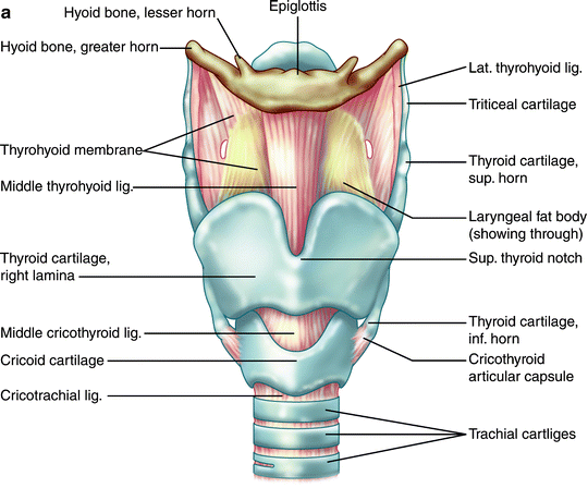
THYROID CARTILAGE 
Features:
- V-shaped in cross-section
- Lamellae
- it consists of right and left lamellae
- posterior parts of lamellae are far apart
- anterior parts of lamellae approach each other
- Laryngeal prominence
- lower parts of right and left lamellae fuse to form this prominence
- they are separated by thyroid notch
- posterior borders are free
- Superior horn
- the lamellae project upwards to form the superior horns
- the superior cornua are connected with greater cornua of hyoid bone through the thyrohyoid ligament
- Inferior horn
- articulates with cricoid cartilage to form cricothyroid joint
- it is connected t cricoid cartilage through conus elasticus
- it is convex in the front and concave in the behind
- oblique line
- the outer surface of the laminae is marked by a slight prominence called oblique line
- extends from the superior thyroid tubercle to the superior cornua
- attachments
- thyrohyoid
- sternohyoid
- thyrophyrangeus part of the inferior constrictor
Attachments :
- the lower border of the inferior cornua
- gives attachments to cricothyroid
- insertion at posterior border connecting superior and inferior cornua
- palatophayrngeus
- salpinophayrangeus
- stylopharyngeus

- inner aspect attachments
- median thyroepiglottic ligament
- thyroepiglottic muscle on both sides
- vestibular fold on both sides
- vocal fold on both sides
- thyroarytenoid
- vocalis muscle on each side
CRICOID CARTILAGE
Features :
- it encircles the larynx below the thyroid cartilage
- it consists of two parts
- the narrow anterior arch
- The inferior cornua of the thyroid cartilage articulate with the side of the cricoid cartilage
- the posterior lamina
- lamina project upwards behind thyroid cartilage and articulates superiorly with the arytenoid cartilage
Attachments :
- the anterior part of the arch - origin to the cricothyroid muscle
- anterolateral part of the arch - lateral cricoarytenoid muscle
- lamina of cricothyroid - posterior cricoarytenoid cartilage

posterior cricoarytenoid muscle is also known as "safety muscle".
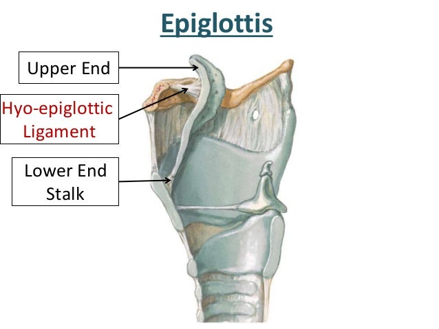
EPIGLOTTIS
Features:
- leaf-shaped
- placed in the anterior wall of the upper part of the larynx
- upper end
- broad and free
- projects behind hyoid bone
- the lower end ( thyroepiglottic ligament)
- attached to the angle between the two laminae of the thyroid cartilage
Attachments:
- anterior surface
- tongue by median glossoepiglottic fold
- To the hyoid bone by the hyoepiglottic ligament
- posterior surface
- covered by mucus membrane and has a tubercle in the lower part

Thyroepiglottic muscle is attached between the thyroid cartilage and margins of the epiglottis.
It keeps the inlet of the larynx patent for breathing.
Aryepiglottic muscle closes inlet during swallowing.
ARYTENOID CARTILAGE

Features:
- two small pyramid-shaped cartilages lying on the upper border of the lamina of the cricoid cartilage
- it has two parts
- the apex
- curved posteromedially and articulates with the corniculate cartilage
- the base
- concave and articulates with the lateral part of the upper border of the cricoid lamella
- the vocal process
- the prolonged process of the lamellae
- the muscular process
- the laterally prolonged process of lamillae
Attachments:
- vocal process
- vocal folds
- vocalis muscles
- above vocal process
- vestibular fold attached
- muscular process
- insertion to posterior cricoarytenoid cartilage
- anterior surface
- lateral cricoarytenoid cartilage
- posterior surface
- transverse arytenoid across the two cartilage

between the base and apex of the arytenoid is the oblique arytenoid which continues as aryepiglottic muscle into two sides of the epiglottis.
The quadrangular or quadrate membrane is attached between arytenoid, epiglottis and thyroid cartilages.
CORNICULATE CARTILAGE
These are two small conical nodules which articulate with the apex of the arytenoid cartilages.
They lie in the posterior parts of the aryepiglottic folds

CUNEIFORM CARTILAGE
These are two small conical nodules that articulate with the apex of the arytenoid cartilages and are directed posteromedially. They lie in the posterior parts of the aryepiglottic folds
LARYNGEAL JOINTS
the joint between different cartilages of the larynx are classified
1. cricothyroid joint
- Synovial joint between inferior cornua of the thyroid cartilage and a side of the cricoid cartilage
- permits rotatory movements at transverse axis through both cricoid cartilage
- this movement permits tension and relaxation of the vocal cords
2. cricoarytenoid joint
- synovial cartilage between the base of arytenoid cartilage as upper lamina of the cricoid cartilage
-permits rotatory movement along the vertical axis
- facilitates abduction and adduction of the vocal cords
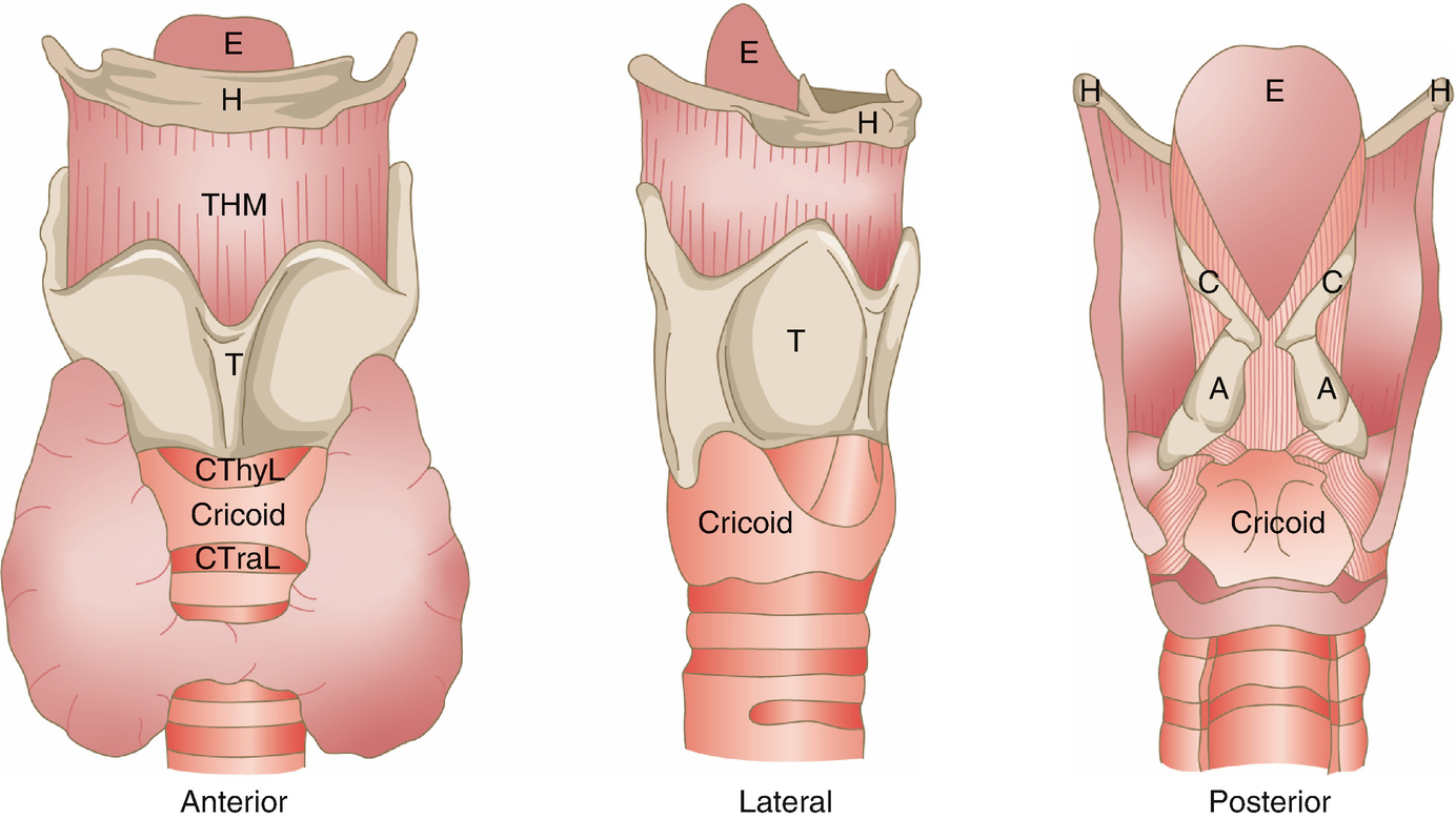
LYRANGEAL LIGAMENTS AND MEMBRANES
Extrinsic:
- thyrohyoid membrane
- connects thyroid cartilage to the hyoid bone
- median and lateral parts are thickened to form the median and lateral thyrohyoid ligaments
- The membrane is pierced by the internal laryngeal nerve, and by the superior laryngeal vessels

- hypoepiglottic ligament
- connects the upper end of the epiglottic ligament
- the upper end of the epiglottic cartilage to the hyoid bone
- cricotracheal ligament
- connects the cricoid cartilage to the upper end of the trachea

Intrinsic muscle :
they are a part of fibroelastic membrane of the larynx.
It is placed outside the mucous membrane.
it consists of a sinus on both sides.
the part of the membrane above the sinus is known as the quadrate membrane
- extends from the arytenoid cartilage to the epiglottis.
- lower free border forms the vestibular fold.
- an upper border forms the aryepiglottic fold
- the part below the sinus is called the conus elasticus.
- extends upwards and medially from the arch of the cricoid cartilage
- The anterior part is thick and is known as the cricothyroid ligament
- The upper free border of the conus elasticus forms the vocal fold
CAVITY OF LARYNX
- Extends from the inlet of the larynx to the lower border of the cricoid cartilage
- The inlet of laynx is oblique and opens into laryngopharynx
- the inlet is bound
- anteriorly by epiglottis
- posteriorly by inter arytenoid fold of mucus
- laterally by aryepiglottic fold
- there are two folds of mucus within the larynx
- upper vestibular fold
- the space between the right and left vestibule is called rima vestibuli
- lower vocal fold
- The space between two vocal folds are called rima glottidus

- the vestibular and vocal folds divide the larynx into three parts
- vestibule - the part above the vestibular fold
- ventricle of larynx - the part between the vestibule and vocal chord
- infraglottis - part below the vocal fold
MUCOUS MEMBRANE OF LARYNX
the mucous membrane is generally lose with the cartilage
it is thin and firm above vocal ligaments and epiglottis. mucous glands are absent over the vocal cords
- Stratified squamous epithelium
- the anterior and upper half of the posterior surface of epiglottis
- the upper part of the aryepiglottic fold
- ciliated columnar epithelium
- rest of the larynx
MUSCULAR ATTACHENTS TO LARYNX
there are 7 muscles attached to the larynx

1. Cricothyroid muscle
origin - lower border and lateral surface of cricoid
insertion - inferior cornua of thyroid cartilage
origin - posterior surface of lamina of cricoid
insertion - posterior muscluar process of arytenoid


3.lateral cricoaryteoid cartilage
origin - upper border of arch of cricoid
insertion - anterior part of muscular process of arytenoid 
4.Transverse arytenoid
unpaired muscle
origin - posterior surface of one arytenoid
insertion - posterior surface of other arytenoid
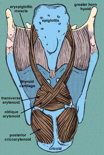
5. oblique arytenoid and aryepiglottic
origin - muscular process of one arytenoid
insertion - the apex of other arytenoid

6.transverse arytenoid or thyroepiglottic
origin - thyroid angle and adjacent cricothyroid ligament
insertion - anterolateral surface of arytenoid cartilage 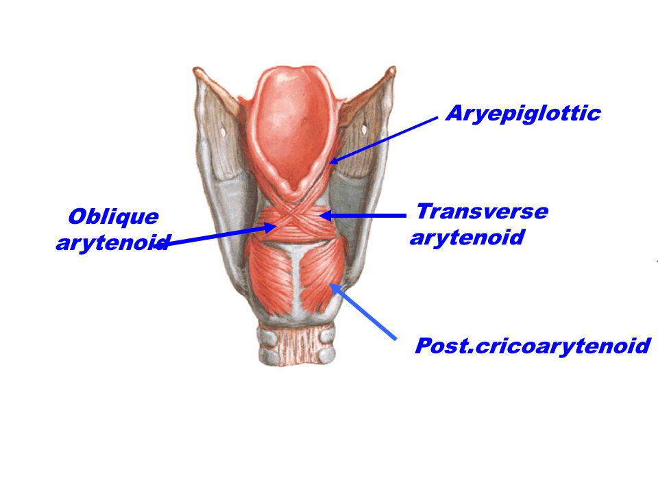
7. vocalis
origin - vocal process of arytenoid cartilage
insertion - vocal ligament at thyroid angle

MOVEMENT OF LARYNX
muscles that pull the muscular process forward and laterally will push the vocal process medially causing adduction
of vocal cords

of vocal cords

muscles that pull the muscular process medially and push the vocal process laterally causes the abduction of vocal cords
muscles that abduct the vocal cord :
- posterior cricoarytenoid
muscles that adduct the vocal cord :
- lateral cricoarytenoid
- transverse arytenoid
- cricothyroid
- thyroarytenoid
muscles that tense the vocal cords:
- cricothyroid
muscles that relax the vocal cord:
- thyroarytenoids
- vocalis
muscles that close the inlet of the larynx :
- oblique arytenoids
- aryepiglottic
muscles that open the inlet of the larynx :
- thyroepiglottis
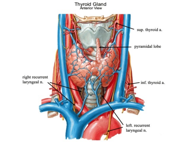
ARTERIAL SUPPLY AND VENOUS DRAINAGE
up to the vocal cords :
- Artery supply - the superior laryngeal artery a branch of the superior thyroid artery
- Venous drainage - superior thyroid vein

below the vocal cords :
- Artery supply - inferior laryngeal artery, a branch of the inferior thyroid artery
- Venous drainage - inferior thyroid vein
NERVE SUPPLY
Motor nerves
- recurrent laryngeal nerve supplies
- posterior cricoarytenoid
- lateral cricoarytenoid
- transverse arytenoid
- oblique arytenoid
- aryepiglottic
- thyroarytenoid
- thyroepiglottic
- external laryngeal nerve
- cricothyroid
Sensory nerves
- internal laryngeal nerve
- mucous membrane up to the level of vocal cord
- recurrent laryngeal nerve
- mucous membrane below the level of vocal cords
LYMPH DRAINAGE
- lymph from above the vocal cord is drained into the anterosuperior group of deep cervical lymph nodes
- lymph from below the vocal cord drain into the posterosuperior part of deep cervical lymph nodes
- a few of them drain into the pharyngeal nodes by piercing cricothyroid

Comments
Post a Comment