MIDDLE PART OF NORMA BASALIS
Introduction :
Norma basalis is the basal view of the skull. considering it as one of the most difficult parts of studying the skull, the following short notes will help you revise and get through it with ease.
Anatomical divisions:
For the sake of understanding, Norma basalis is divided into three parts
1.Anterior part of Norma basalis
2. Middle part of Norma basalis
3. posterior part of Norma basalis
MIDDLE PART OF NORMA BASALIS
borders of the middle part of Norma basalis :
anterior: horizontal plates of the palatine
posterior : anterior part of foramen magnum
lateral: styloid and mastoid process
Middle landmarks of Norma basalis :
- greater wing of the sphenoid
- petrous part of the temporal bone
- mastoid process
- styloid process
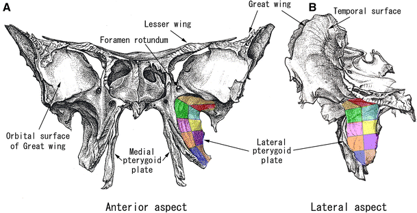
Pterygoid process of the sphenoid

Contents:
1.pterygoid Hamulus
2.medial pterygoid plate
3.lateral pterygoid plate
4.scaphoid fossa
5.vaginal process
6.palatovaginal canal
7.vomerovaginal canal
8.emissary sphenoidal foramen
9.spine of sphenoid
Muscular attachment:
- medial pterygoid muscle (attached to medial pterygoid plate)
- lateral pterygoid muscle (attached to lateral pterygoid plate)
- tensor veli palatine
- phyrangobasilar ligament (medial pterygoid plate)
- superior constrictor (originates at pterygoid Hamulus)
- pterygomandibular raphe (attached to pterygoid hamulus)
- muscles attached to the spine of sphenoid
- spinomandiular ligament
- anterior ligament of malleus
- pterygospinous ligament
canal contents:
palatovaginal canal: pterygopalatine ganglion
vomerovaginal canal: pharyngeal branch of the pterygopalatine ganglion
GREATER WING OF SPHENOID:
 Contents:
- foramen ovale
- foramen rotundum
- foramen spinosum
- emissary sphenoidal foramen: emissary vein connecting cavernous sinus and pterygoid plexus
muscular attachment:
1.sphenomandibular ligament
2.temporalis
3.cartilaginous part of the auditory tube (attached to sulcus tube)
4.upper head of lateral pterygoid

Foramen of the greater wing of sphenoid :
foramen ovale:
- maxillary artery
- accessory meningeal artery
- lesser petrosal nerve
- emissary's vein
foramen spinosum:
- middle meningeal artery
- the posterior trunk of the middle meningeal vein
- meningeal branch of the mandibular nerve
PETROUS PART OF TEMPORAL BONE
Contents:
- foramen lacerum
- jugular foramen
- carotid canal
- inferior tympanic canalicus (canaliculus innominatus)
muscle: levator veli palatini
Foramen contents:
foramen lacerum contains:
- Emissary's vein
- Middle meningeal branch of the accessory pharyngeal artery
structures passing above foramen lacerum:
- Internal carotid artery
- deep petrosal and lesser petrosal nerve surrounds internal carotid artery to form nerve of pterygoid canal
Carotid canal contains:
- Internal carotid artery
- sympathetic plexus

Jugular foramen contains:
- inferior petrosal sinus
- glossopharyngeal nerve
- vagus nerve
- the cranial part of accessory
- sigmoidal sinus
- meningeal branch of the occipital and ascending pharyngeal nerve
Muscular attachment of petrous temporal
1. Levator veli palatine
MASTOID PROCESS:
Foramen :
1.mastoid canaliculus : auricular branch of vagus nerve
2. mastoid foramen : (rare) transmits emissary vein to transverse sinus and a small branch of occipital artery to dura matter
Muscular attachments:
- longissimus capitus
- splenius capitus
- sternomastoid muscle
- posterior belly of digastric

STYLOID PROCESS
present in front of the mastoid process
Foramens:
stylomastoid foramen: transmits stylomastoid artery and facial nerve
Muscular attachments:
- stylomandibular ligament
- styloglossus muscle
- stylophyrangeus muscle
- stylohyoid muscle
- stylohyoid ligament


Comments
Post a Comment