Thyroid cartilage
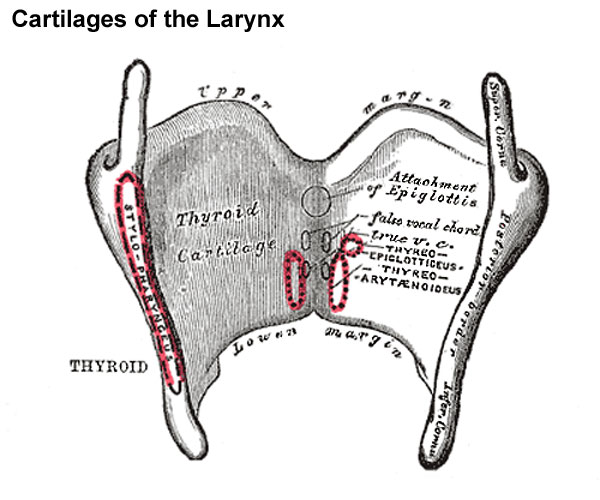
THYROID CARTILAGE

Features:
- V-shaped in cross-section
- Lamellae
- it consists of right and left lamellae
- posterior parts of lamellae are far apart
- anterior parts of lamellae approach each other
- Laryngeal prominence
- lower parts of right and left lamellae fuse to form this prominence
- they are separated by thyroid notch
- posterior borders are free
- Superior horn
- the lamellae project upwards to form the superior horns
- the superior cornua are connected with greater cornua of hyoid bone through thyrohyoid ligament
- Inferior horn
- articulates with cricoid cartilage to form cricothyroid joint
- it is connected to cricoid cartilage through conus elasticus
- it is convex in the front and concave in the behind
- oblique line
- the outer surface of the laminae is marked by a slight prominence called oblique line
- extends from the superior thyroid tubercle to the superior cornua
- attachments
- thyrohyoid
- sternohyoid
- thyrophyrangeus part of the inferior constrictor
Attachments :
- the lower border of the inferior cornua
- gives attachments to cricothyroid
- insertion at posterior border connecting superior and inferior cornua
- palatophayrngeus
- salpinophayrangeus
- stylopharyngeus

- inner aspect attachments
- median thyroepiglottic ligament
- thyroepiglottic muscle on both sides
- vestibular fold on both sides
- vocal fold on both sides
- thyroarytenoid
- vocalis muscle on each side


Comments
Post a Comment