visual pathway
PATHWAY
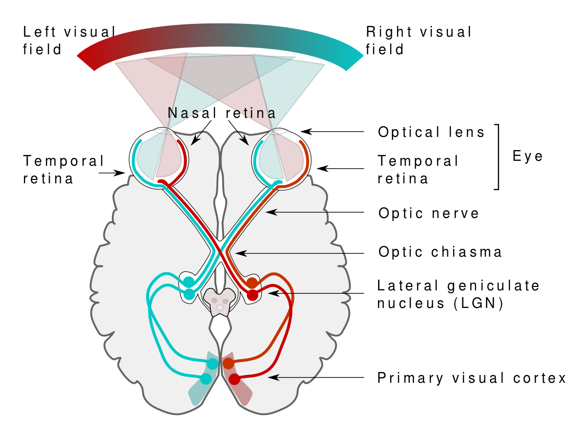
Visual pathway relays signal in the following way:
Receptors receive signals
They synapse with bipolar cells
Bipolar cells synapse with ganglion cells
Ganglion cells from the optic nerve
Fibres of the optic nerve cross over each other partially at the optic chiasma
The nasal half of the retina crosses over at optic chiasma (retinal half stays intact)
After crossing over the optic track is formed
The optic track reaches the lateral geniculate body
From the lateral geniculate body, it proceeds as optic fibres
Optic fibres end in the primary visual cortex
Note: the optic track is formed by optic tracks from the opposite side and the temporal track of the same side
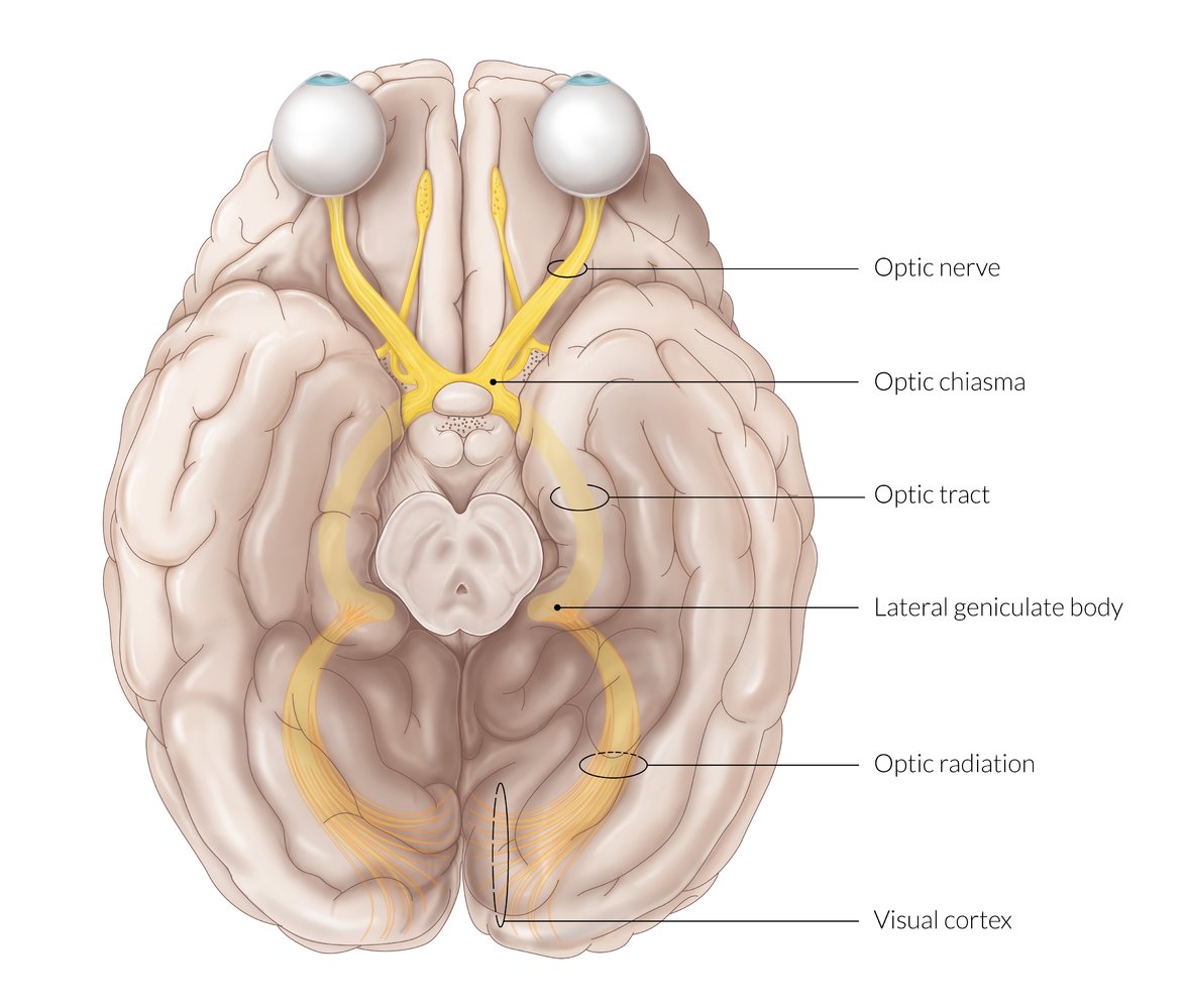
In short :
1. Optic nerve
2. Optic chiasma
3. Optic tract
4. Lateral geniculate body
5. Optic radiation
6. Visual cortex.

Optic nerve
The optic nerve is formed by axons of ganglionic cells
Leaves eye through optic disk
The temporal part of the optic nerve carries impulse from the nasal half
The nasal part of optic nerve carries impulse form temporal half
Optic chiasma
Medial fibres of each optic nerve cross the midline and join the uncrossed lateral fibres of the opposite side, to form optic track
The cross over area is called optic chiasma
Optic tract
LATERAL GENICULATE BODY
The optic track brings signals from eyes and ends in the lateral geniculate body
From the lateral geniculate body the neurons process as optic fibres and end in the visual cortex

The lateral geniculate body has 6 layers named from 1 to 6.
Layers 1, 4 and 6 receive inputs from the medial half of the eye
Layers 2, 3 and 5 receive inputs from the lateral half of the eye
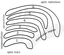
MAGNOCELLULAR LAYER
Layers 1 and 2 contain large neurons called magnocellular layers
These cells are called M cells (Y type retinal ganglion cell)
They project to the magnocellular layer of LGB
They transmit black and white visual signals
PARVOCELLULAR LAYER
Layers 3 to 6 have small to medium-sized nerves called the parvocellular layer
X type cells and P-type cells of retinal ganglion project to this layer
The velocity of transmission is slow


Comments
Post a Comment