Temporomandibular joint
Temporomandibular joint
It is a synovial joint of condylar variety
It connect the mandible with the caverna
Relations
Anterior - lateral pterygoid ,mesenteric vessels and parotid gland
Posterior - superficial temporal vessels and auriculotemporal nerve
Superior - middle meningeal vessels and middle cranial fossa
Inferior - maxillary artery and vein
Lateral - skin, fascia, parotid gland, temporal branches of the facial nerve
Blood supply - superficial temporal and maxillary artery
Nerve supply - auriculotemporal nerve and maxillary nerve.
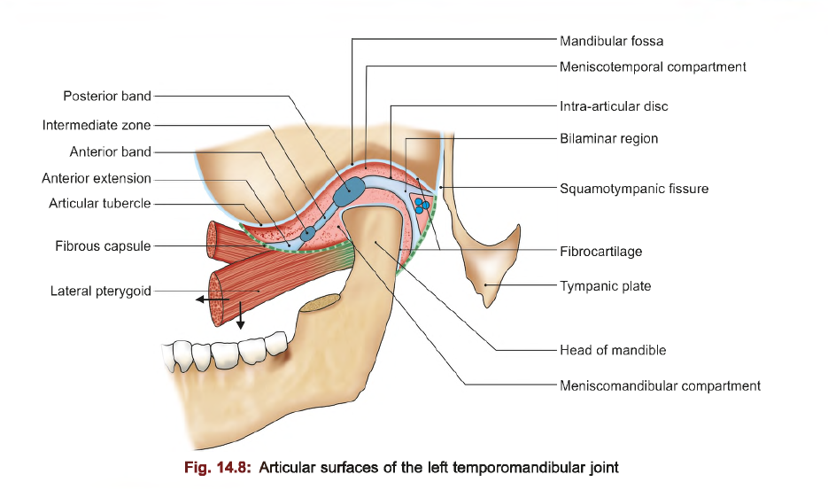
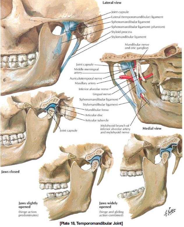
there are 5 ligaments attached to the temporomandibular joint
Fibrous capsule
Lined by synovial membrane.
Attached to the auricular tubercle
sqamotympanic fissure
Temporomandibular ligament
Attached to the auricular tubercle
head of mandible
Strengthens the lateral part
Spheinomandibular ligament
Attached to the spine of the sphenoid and lingua of the mandibular foramen
It is a remnant of Meckel's cartilage(embryonic cartilage that contributes to the formation of mandible)
Pierced by the nerve to mylohyoid in the inferior segment
Stylomandibular ligament
Attached to the styloid process and the posterior ramus of the mandible
It separates the parotid and submandibular region
Pterygomandibular ligament
Attached to the pterygoid hamulus and the inner aspect of the mandible near the third molar

Joint capsule:
Contains 3 parts
Meniscotemporal compartment - upper part, permits gliding movement
Meniscomandibular compartment- lower compartment, permits both gliding and rotatory movement
Articular disc-
lies in between the upper two compartment
Fibrous in nature
Provides cushioning for the head of the mandible
Prevents friction in the articulating surface and lowed
It consists of several parts
1. bilaminar region
2. posterior thick region.
3.Intermediate band.
4.Anterior thick region.
5. Anterior region
Synovium:
Tmj is lined with synovium and synovial fluid
The synovial fluid is a filtrate of plasma and consist of hyaluronic acid
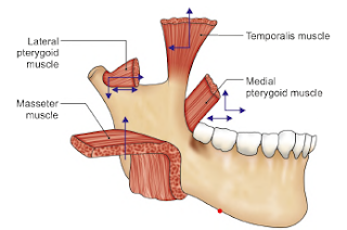
Muscles involved in mandibular movements:
Depression- lateral pterygoid digastric geniohyoid and mylohyoid
Elevation- masseter, medial pterygoid, temporalis
Protrusion - lateral and medial pterygoid
Retraction - horizontal fibres of temporalis and masseter


Comments
Post a Comment