Oral cavity
Oral cavity
Vestibule
Oral fissure
Oral cavity proper
VESTIBULE:
It communicates with oral fissure
The parotid duct opens in the vestibular region beside the second maxillary molar
The entire vestibule is lined by mucous membrane
Several glands open in the vestibule
The oral vestibule connects with the gums through the frenula of the lips
Clinical feature: in the case of parotid calculi the dye is injected through the parotid duct in the vestibule
CHEEKS:
Is fleshy flap of covering on both sides of the oral cavity
The surface between the oral fissure and the cheek is called a nasolabial sulcus
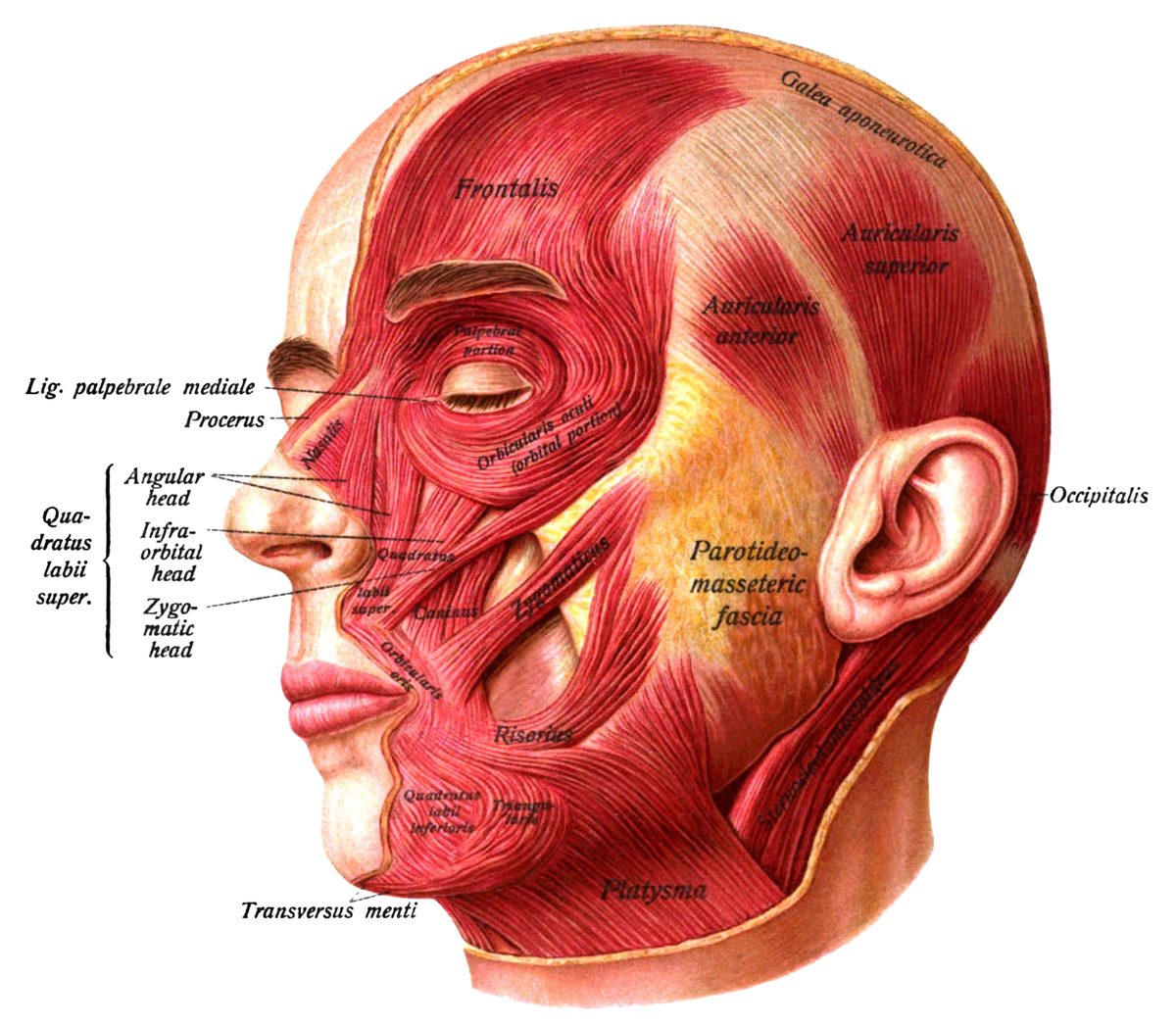
1. Skin
2. Superficial fascia
3. Fatty pads of the cheek( infraorbital fat pads, lateral orbital fatty pads and nasolabial fat pads)

4. Parotid duct
Deep to the masseter and slightly superficial to the buccinator buccal pad of fat is present, it drains the parotid gland secreation.
it is innervated by the facial and trigeminal nerve
- Frenulum
- Sublingual papilla (with sublingual salivary duct opening)
- Sublingual mucous fold
Posterior relations:
- tongue
- oropharyngeal isthmus

1. free gum
2.attached part which continues with periodontal
membrane

Bones:
Posterior one third - horizontal wall of the palatine.
Surfaces:
Posterior surface-soft palate
Superior surface - floor of the nasal cavity
Inferior surface - the roof of the oral cavity
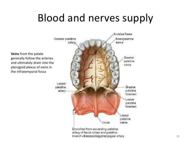
VESSELS OF HARD PALATE:
Arteries - a great palatine branch of the maxillary artery
Vein - pterygoid plexus of veins
Nerves - great palatine and lesser palatine branch of the maxillary nerve
Lymph - upper deep cervical nodes
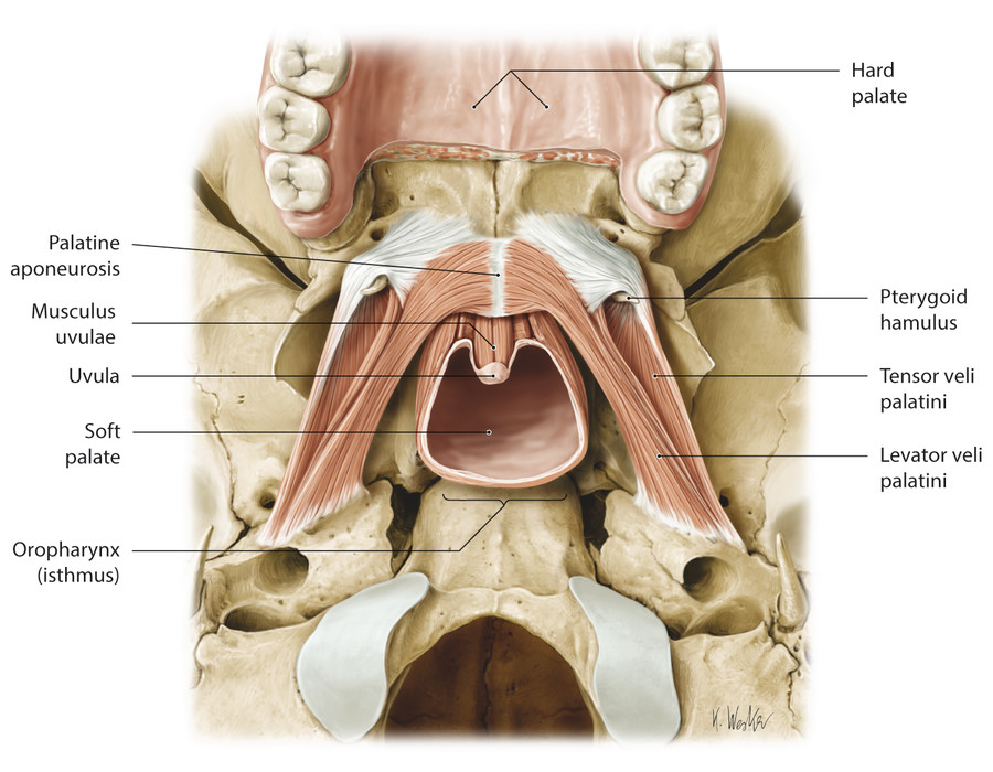
SOFT PALATE
Anterior relation - marked by median raphe
Posterior surface - nasal cavity floor
Superior surface - hard palate
Inferior border - uvula and palatoglossal arch
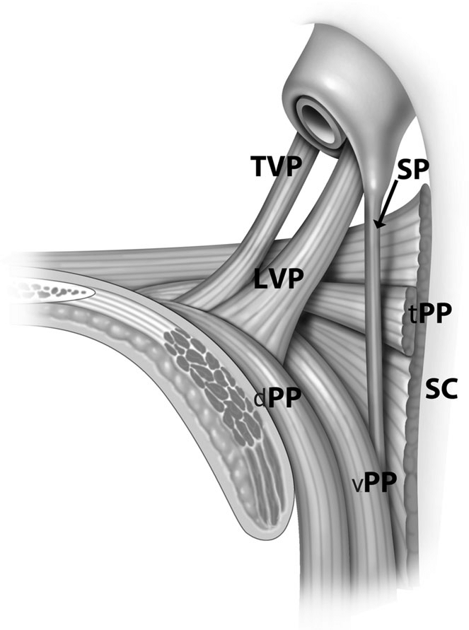
CONTENTS OF SOFT PALATINE OF SOFT PALATE :
Palantine aponeurosis (tendon of tensor villi palantine)
Levator villi palantine and palatophyryngeus (lies superior to the palantine aponeurosis)
NERVE SUPPLY :
MOTOR NERVE :
all five muscles except tensor villi palatine are supplied by the pharyngeal plexus.
Tensor villi palatine is supplied by the mandibular nerve
SENSORY NERVE:
The middle and posterior lesser palatine nerve
Glossopharyngeal nerve
GUSTATORY NERVE:
Comes from the nucleus of tractus solitarius
Supplies through lesser palatine nerve
SECRETOMOTOR PATHWAY:
From superior salivatory nucleus
MUSCLES OF SOFT PALATE


PASSAVANT RIDGE :
The mucosal ridge formed by the superior constrictor and palatopharyngeal muscle
Covers the pharyngeal isthmus between the nasopharynx and oropharynx
It is a U shaped loop of mucosae
Aka palatopharyngeal sphincter
Functions of soft palate :
Separates oro and nasopharynx
Forms passavent ridge
Control of voice using pharyngeal isthmus


Comments
Post a Comment